Sources & credits
Sources & licences
Data, software and font licences for this site.
Project images. The project images come from model outputs, recordings and product renders. Their data sources and licence terms are listed below.
Connectivity, skeletons & body replay
Fly circuit study
The interactive fly explorer combines saved paired MN9 output with a bounded derivative of published FlyWire v630 connectivity. Neuron IDs, connection counts and model signs are retained. The connections view uses an original graph layout. The anatomy view adds real reconstructed skeletons for the same source IDs, in their shared coordinate frame, with documented branch simplification and unit conversion. The complete raw connectome is not distributed. Paired replay provenance, network selection and anatomy processing and source hashes document these parts.
The separate body illustration includes geometry from FlyBody, under Apache 2.0, and local Three.js 0.160.0, MIT. A separate simulation archive drives an assumed, uncalibrated movement mapping; it is not the paired sensitivity experiment and does not establish physical feeding. The body and neuron skeletons have no verified registration. Body sources and processing.
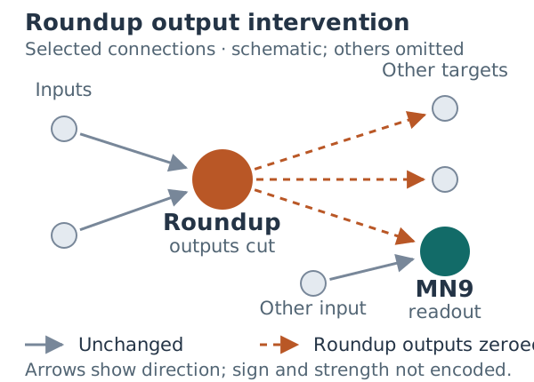
The focused circuit diagram selects directed connections from the model export. Dashed outgoing links mark the Roundup-output intervention. Selection, attribution and source fingerprints.
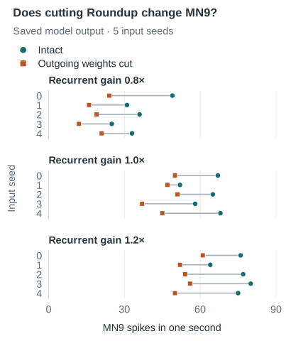
The homepage recorded replay and poster are captured from the body replay, with simultaneous whole-fly and mouth-detail views. The compact paired-count chart redraws the original saved experiment. Their distinct sources, capture window and file hashes are documented in media provenance; the replay is not animal footage or paired-study movement.
The model code is MIT-licensed. FlyWire-derived connectivity and annotations retain CC BY-NC 4.0, following the FlyWire published-data principles. This research display selects a small subset and adds a layout; it does not change the source connection counts or annotations. Credit: the FlyWire Consortium; Dorkenwald et al. (2024); Schlegel et al. (2024); synapse detection by Buhmann et al. (2021) and Heinrich et al. (2018); transmitter predictions by Eckstein, Bates et al. (2024). The displayed sign is the source model’s assigned sign, not a new measurement of transmitter identity.
The research figures show saved outputs of exploratory comparisons, not figures copied from the source papers. Their aggregate tables and source hashes are in the evidence download. Raw recordings and the complete connectome files are not distributed here.
- Fly circuit: Shiu et al. (2024) and the published Drosophila brain model at commit 91bdd1e7. This portfolio's sensitivity study is not a biological validation or full paper replication.
Saved synthetic observations
Rare-error study
This independent methods study uses synthetic data with known generating factors. The controls display saved results; they do not run a new fit. Biological recordings have not been evaluated.
Method: Kobak et al. (2016), using the pinned Machens Lab Python implementation.
- Interactive study
- Saved metrics (CSV)
- Study configuration
- Provenance & file hashes
- Original frozen figure
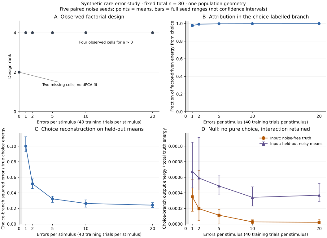
The downloads contain the saved results and configuration; the study’s analysis code is not included.
One population geometry, fixed signal strengths and one regularizer limit the conclusion. These curves do not establish a universal minimum error count or causal choice coding.
Population-average atlas reference
MorphBrain
The GW21–36 playback uses weekly white-matter surfaces derived from the Developing Human Connectome Project spatio-temporal surface atlas of the fetal brain. The source atlas is a population-average reference released under the CC0 1.0 Public Domain Dedication; it is not subject MRI and is not the molecular model developing.
Source: Karolis et al., Developing Human Connectome Project spatio-temporal surface atlas of the fetal brain.
Recording & source meshes
Model Morpher
The recorded DG-to-root example uses Allen Mouse Brain Common Coordinate Framework meshes delivered by
BrainGlobe as allen_mouse_25um v1.2. Image/data credit: Allen Institute. This is a
noncommercial research demonstration.
Bundled dependencies
Software & fonts
The MorphBrain browser build includes React, three.js, React Three Fiber, Drei, Zustand and their transitive open-source dependencies. The geometry decoder is Google Draco, licensed under Apache-2.0.
The portfolio shell uses self-hosted Source Sans 3 for body and interface text and DM Mono for figure and source labels. Spectral is also distributed in the bundled assets. All three are licensed under the SIL Open Font License 1.1.
Package-level copyright notices and licence texts are preserved in the complete third-party notice. The shared licence texts are also available under the licences directory.
The separate portfolio design draft also uses Caveat, self-hosted under the SIL Open Font License 1.1. Its decorative fly reuses the existing FlyBody source geometry (Apache 2.0); the translucent material and scroll-linked camera are presentation choices, not measured neural activity or a behavioural simulation.
Interface motion
GSAP 3.15.1 and its ScrollTrigger plugin are self-hosted for interface motion. Their copyright and standard licence declaration are preserved in the distributed GSAP notice. Lenis 1.3.11 is self-hosted for wheel smoothing under the MIT licence. No third-party script server is contacted.
Self-hosted presentation
The portfolio shell
The page you are reading contacts no other host. There are no CDN resources, no analytics and no
external font services, and the Content-Security-Policy declared in its <head>
forbids them — fonts, styles, scripts and media may only be fetched from this domain.
The interface uses self-hosted styles, scripts and fonts, with the motion libraries noted above. Font licences are reproduced in full alongside the other notices.
Portfolio-specific writing, media and presentation are not offered under an open-source licence. Each third-party component remains governed by its own terms.