Jason Wang
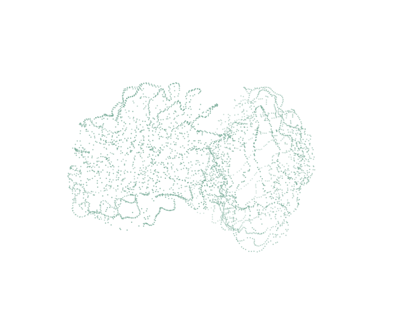
Points sampled from a week-36 fetal white-matter atlas surface, drawn as a static outline of the brain

Jason Wang

Neuroscience student at UCL. I build models, signal analyses and scientific software for looking at brains.

Interactive atlas surface study

Move over the surface, drag to turn it, or focus it and use the arrow keys. Home resets the view.

4,096 points

4,096 vertices sampled from the week-36 population-average white-matter surface of the dHCP fetal surface atlas (CC0).

The way points glow and part around your cursor is interface artwork. It is not neural activity, biological dynamics or development, and the display orientation is not anatomically validated.

Sampling and source record (JSON)

Selected work.

Explore the things I build to understand brains, shapes and systems.

Start exploring

Geometry / Python

Between
forms.

Model Morpher
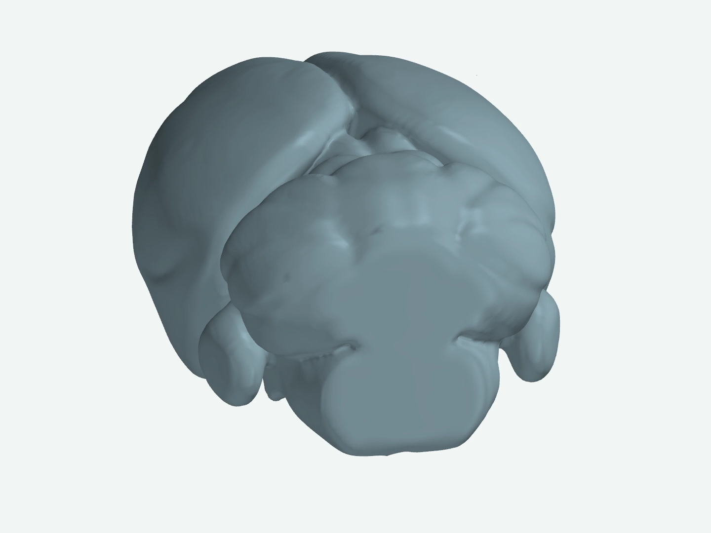
Static preview from the saved Model Morpher rendering; this poster does not indicate the selected interactive frame.

Static saved rendering. The full sequence is in the recording.

Geometric interpolation, not anatomical correspondence or development. Inspect shows three exact saved surfaces; the recording shows the whole sequence.

I built a Python tool that generates the shapes in between two meshes, even when their topology differs, by blending their signed distance fields. Here it runs from the dentate gyrus to the whole mouse brain, using the Allen Mouse CCF through BrainGlobe.
Watch the full sequence 5.5 s
Full sequence, 5.5 s: dentate gyrus to whole mouse brain. Poster is a still from the recording at 2.67 s.

Morphliner

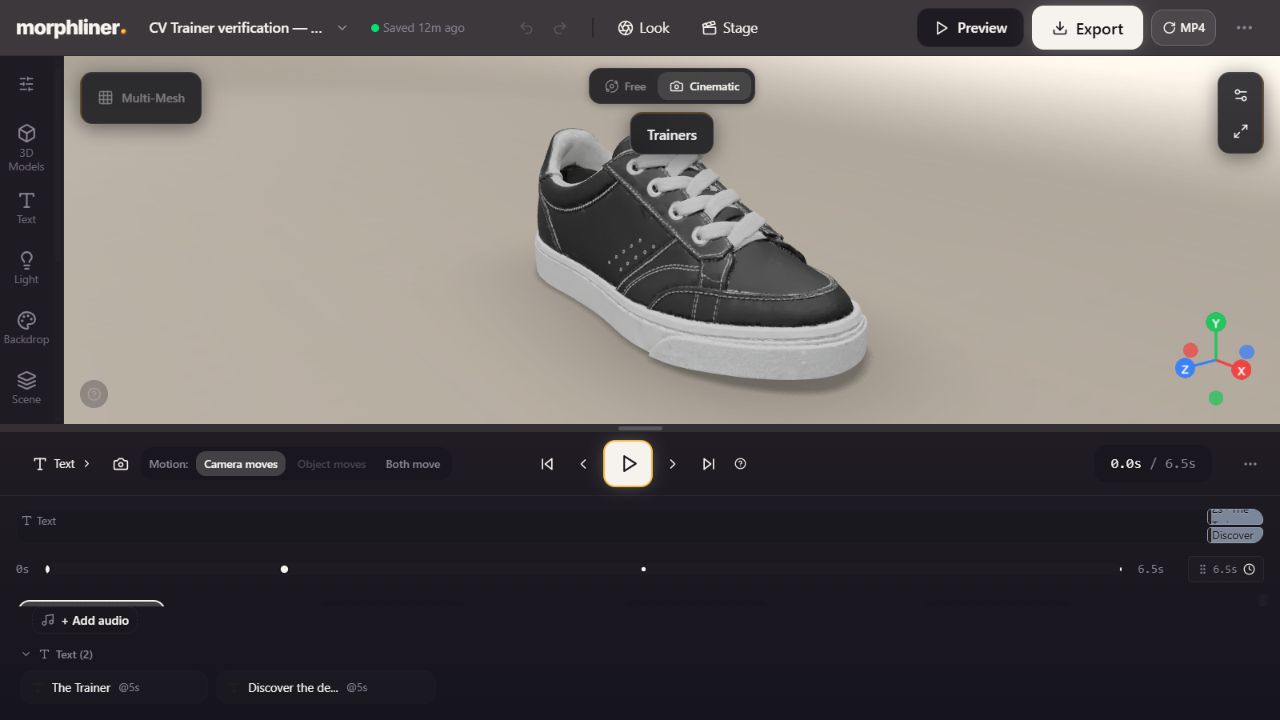
A 3D editor in the browser. You place camera keyframes on a scene, and it exports the move as a video file. The film here is one of those exports.

Exported film, 6.5 s: the Trainer camera sequence.
Inside the editor Screenshot of the Morphliner editor in a browser, with a 3D scene and camera keyframes.

The editor view is a screenshot, not a live editing session.

EEG / team BCI project

When does
the signal
become useful?

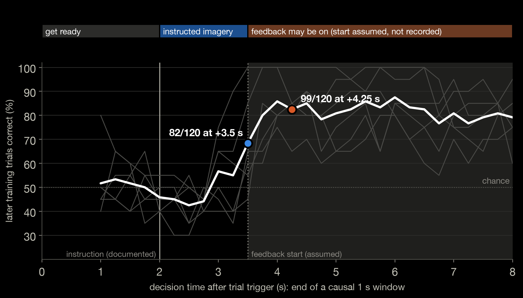
When does EEG imagery become decodable?

Team figure: later training trials correct, in percent, against decision time from 0 to 8 s. The line stays near 50% chance until about 3 s, then rises to around 80%. Marked points: 82/120 at +3.5 s and 99/120 at +4.25 s.
The team's original timing figureEnlarge figure
  1. Get ready, 0–2 s
  2. Imagery, 2–3.5 s
  3. Feedback may be on from 3.5 s (assumed)

The 1 s window 2.5–3.5 s ends where feedback is assumed to begin.The 1 s window 3.25–4.25 s overlaps the assumed feedback by 0.75 s.

Both scores are from later training trials we had already inspected, with a separate fit for each shifted 1 s window. Not a held-out gain. 3 people, 6 sessions.

A team BCI project. I contributed evaluation pull requests and obtained and relayed the organisers' scoring rules. The figure is the team's, unchanged.

Selector result

A selector choosing settings from the 60 calibration trials per session scored 75/120, below 82/120 for fixed CSP.

Whole-brain model / fly circuit

A connection.
A consequence.

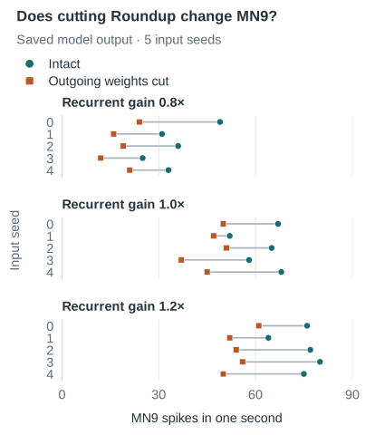
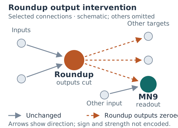
In a published whole-brain fly model implementation, cutting Roundup's outputs reduced MN9 spiking in all 15 paired comparisons, 30 simulations in total.

A model simulation, not feeding behaviour and not an animal experiment.

  • Roundup
  • MN9
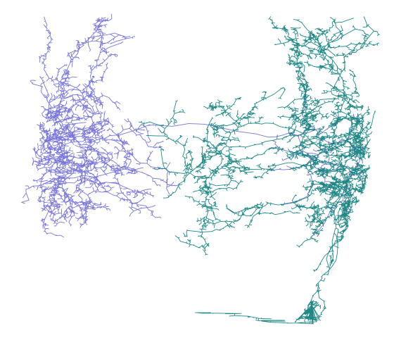
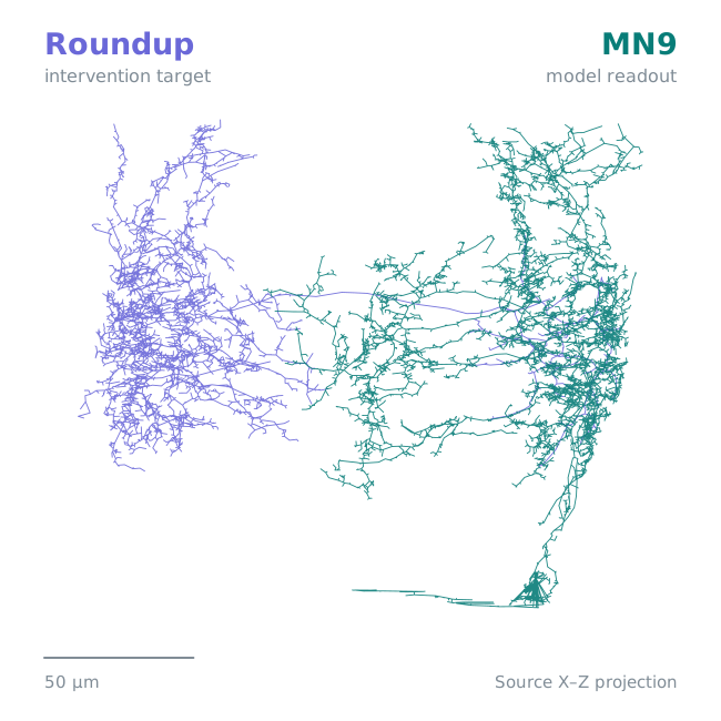
Reconstructed skeletons of Roundup, in violet, and MN9, in teal, with every reconstructed edge drawn in a common source X–Z projection.
50 µmSource X-Z projection
Roundup and MN9 reconstructed neurons from FlyWire v630. Static source X-Z projection showing shape, not activity. Sources and scope.

MN9 spikes in one simulated second

  • Roundup intact
  • Roundup outputs cut
MN9 spiking with Roundup intact and with its outputs cut, for all 15 paired comparisons. Spiking is lower with the outputs cut in every pair.

Each row is one pair: the same input seed (0–4) run twice, once intact and once with all of Roundup's outgoing weights set to zero and its incoming weights retained. Input seeds are simulation inputs, not animals. This is one model under one exploratory input regime, not biological validation. Full counts are in the table below.
All 15 paired comparisons
MN9 spikes in one simulated second, Roundup intact and with its outputs cut.
Recurrent gainInput seedIntactOutputs cut
0.804924
0.813116
0.823619
0.832512
0.843321
1.006750
1.015247
1.026551
1.035837
1.046845
1.207661
1.216452
1.227754
1.238056
1.247550
Neuron sources and scope

Download the labelled neuron figure

Exact saved Roundup (720575940623211725) and MN9 (720575940660219265) skeletons in shared source coordinates, with the existing 0.5 µm display simplification. Every saved edge is retained. Source axes are not verified anatomical directions; no registration to the fly body is claimed. This is morphology, not activity, synapses or a signal route.

FlyWire Consortium / Schlegel et al. and Dorkenwald et al.; skeletons hosted by MRC LMB. FlyWire materialization 630, CC BY-NC 4.0. Source details in the explorer.

Original figures
Separate illustrative replay 9.6 s
Illustrative, uncalibrated body movement from a separate archive, 9.6 s. Not synchronised with the 15 paired comparisons.

Patterning / interactive model

Change the source.
Explore the pattern.

MorphBrain

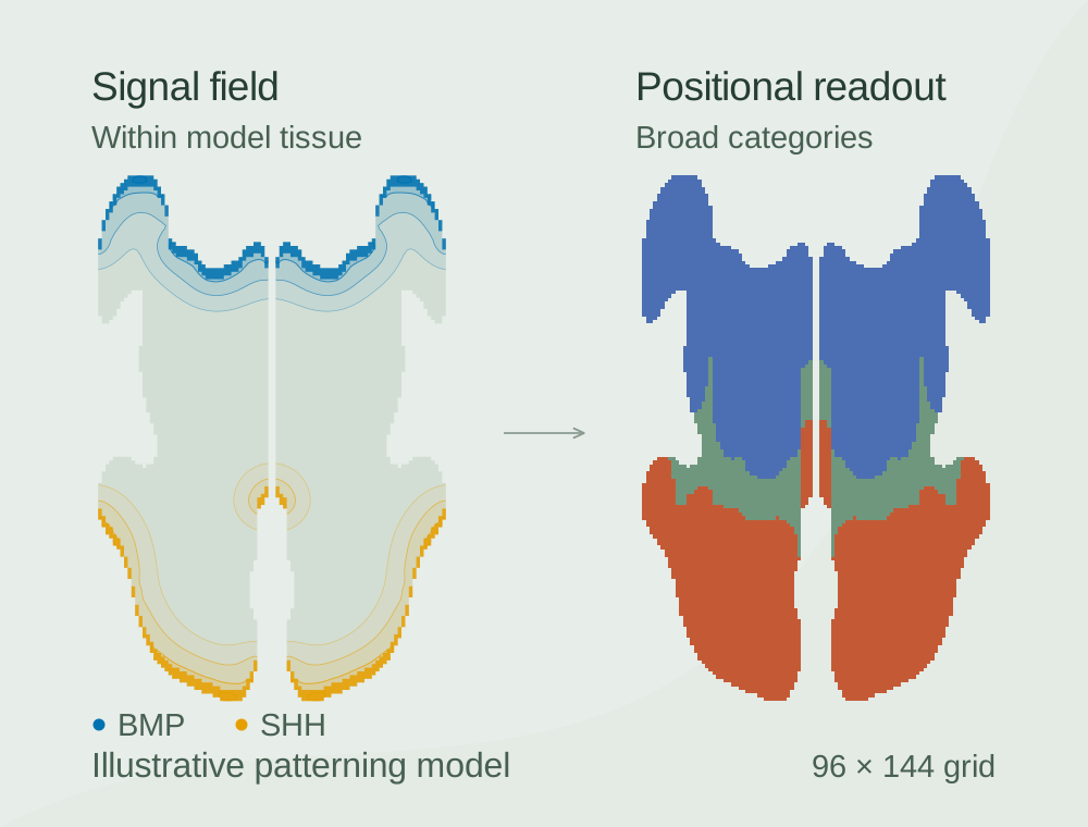
Contours of the SHH and BMP fields in the model tissue, beside the dorsal, intermediate and ventral identities computed from the same state.
One saved state: field contours and the regional identities computed from them.

A patterning model that runs in the browser. Move or remove the SHH and BMP sources, and the regional identities it assigns change.

Illustrative patterning; parameters not calibrated. The folding aim remains open.

Scroll to inspect the full-size figure. Keyboard: Tab to the figure, then use arrow keys.

Team figure: later training trials correct against decision time, 0 to 8 s, with 82/120 marked at +3.5 s and 99/120 at +4.25 s.

About

  • StudyingNeuroscience, UCL
  • ResearchUndergraduate research assistant, UCL
  • Based inLondon

I study neuroscience at UCL and build tools for exploring brains and data. As an undergraduate research assistant with Professor Lorenzo Fabrizi in UCL’s Medawar Pain and Somatosensory Labs, I work on EEG source modelling: relating models of brain activity to signals measured at the scalp.

Also on the full work page: a synthetic study of separating stimulus and choice signals when errors are rare, and the fly input-timing comparison.

I’m interested in paid internships, scientific software work and research opportunities in brain modelling, computational neuroscience and machine learning.